fMRI

Moderátor: Lucid Dreaming Project

Uživatelský avatar
ATA
Příspěvky: 1600
Registrován: stř, 28. kvě 2008, 18:55
Bydliště: Litovel
Kontaktovat uživatele:

fMRI

Příspěvekod ATA » čtv, 27. čer 2013, 03:39

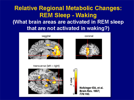
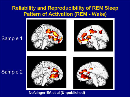
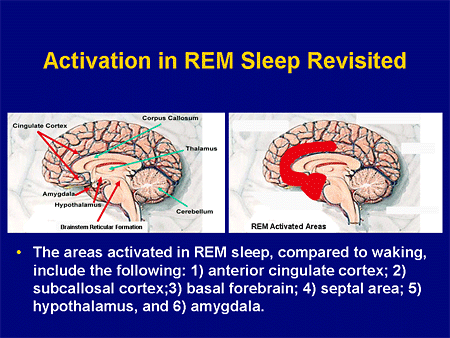
Consistent with the hypothesis that REMs share the brain systems and mechanisms with waking eye movements and are visually-targeted saccades, we found REM-locked activation in the primary visual cortex, thalamic reticular nucleus (TRN), 'visual claustrum', retrosplenial cortex (RSC, only on the right hemisphere), fusiform gyrus, anterior cingulate cortex, and the oculomotor circuit that controls awake saccadic eye movements (and subserves awake visuospatial attention). Unexpectedly, robust activation also occurred in non-visual sensory cortices, motor cortex, language areas, and the ascending reticular activating system, including basal forebrain, the major source of cholinergic input to the entire cortex. REM-associated activation of these areas, especially non-visual primary sensory cortices, TRN and claustrum, parallels findings from waking studies on the interactions between multiple sensory data, and their 'binding' into a unified percept, suggesting that these mechanisms are also shared in waking and dreaming and that the sharing goes beyond the expected visual scanning mechanisms. Surprisingly, REMs were associated with a decrease in signal in specific periventricular subregions, matching the distribution of the serotonergic supraependymal plexus. REMs might serve as a useful task-free probe into major brain systems for functional brain imaging.

REM-locked activation data (při pohybu očí ve snu)

Obrázek
peaks
Obrázek
Obrázek
Obrázek

TRN may control the “attentional search light” during visual scanning
RSC is responsive to scene layout
The anterior cingulate may play a similar role in dreaming, selecting visual targets for scanning under affective and cognitive influence.

Waking imagery studies [

scanpaths (repetitive sequences of fixations and saccades) during visual perception are highly correlated to those during visual imagery of the same visual object; they presented evidence that oculomotor information is stored together with the visual representation and is used as a spatial index for the proper arrangement of parts of an image during image generation. Likewise, in dreaming REMs may reenact the scanpath eye movements in waking visual perception of the same object or scene and retrieve the visual representation encoded together with the scanpath.

Spatially distributed, synchronous gamma oscillations have been proposed to mediate ‘binding’ of multiple sensory data into a unified object representation both in wakefulness [

REM-locked activation was the most (and extraordinarily) robust at the primary visual cortex and TRN. We speculate that TRN plays a key role in the ‘priming’ (preparation in anticipation) of non-visual sensory and motor cortices induced by internal or external visual stimuli, linked to REMs in dreaming or to scanning eye movements in waking, perhaps for faster detection and response. All axons passing between thalamus and cortex traverse TRN where many give off collaterals; TRN has distinct sectors (visual, auditory, somatosensory, motor);

The basal forebrain cholinergic system can induce 40-Hz synchronization and regional enhancement of cortical sensory processing

http://www.ncbi.nlm.nih.gov/pmc/articles/PMC2753360/
Help! I'm a multidimensional being trapped in a linear time-space continuum!

Uživatelský avatar
ATA
Příspěvky: 1600
Registrován: stř, 28. kvě 2008, 18:55
Bydliště: Litovel
Kontaktovat uživatele:

Re: fMRI

Příspěvekod ATA » čtv, 27. čer 2013, 14:07

Obrázek
Help! I'm a multidimensional being trapped in a linear time-space continuum!

Uživatelský avatar
ATA
Příspěvky: 1600
Registrován: stř, 28. kvě 2008, 18:55
Bydliště: Litovel
Kontaktovat uživatele:

Re: fMRI

Příspěvekod ATA » čtv, 27. čer 2013, 14:15

Obrázek
phasic REM sleep periods with high number of rapid eye movements, high cortico-thalamic activity (red) is generated in the brain.

Obrázek
During wakefulness, acoustic stimulation elicits activation of the auditory cortex (red). Similar stimulation during light NREM sleep stage 2 evokes transient downregulation of widespread cortical areas (blue), supposedly a sleep protective mechanism
Help! I'm a multidimensional being trapped in a linear time-space continuum!

Uživatelský avatar
ATA
Příspěvky: 1600
Registrován: stř, 28. kvě 2008, 18:55
Bydliště: Litovel
Kontaktovat uživatele:

Re: fMRI

Příspěvekod ATA » čtv, 27. čer 2013, 16:08

NREM

Obrázek
areas involved in the processing of memory traces such as para-hippocampal areas, the study might point to the potential functions of sleep, in particular the increasingly well-defined role of sleep in memory consolidation. Finally, the activation of areas such as the brainstem, usually associated with arousal and waking, might reveal these oscillations of non-REM sleep as ‘micro-wake’ periods allowing the brain to fulfil crucial and active functions, even during the deepest stages of sleep.
Help! I'm a multidimensional being trapped in a linear time-space continuum!

Uživatelský avatar
ATA
Příspěvky: 1600
Registrován: stř, 28. kvě 2008, 18:55
Bydliště: Litovel
Kontaktovat uživatele:

Re: fMRI

Příspěvekod ATA » čtv, 27. čer 2013, 17:05

Obrázek
Obrázek
Obrázek
Help! I'm a multidimensional being trapped in a linear time-space continuum!

Uživatelský avatar
ATA
Příspěvky: 1600
Registrován: stř, 28. kvě 2008, 18:55
Bydliště: Litovel
Kontaktovat uživatele:

Re: fMRI

Příspěvekod ATA » čtv, 27. čer 2013, 17:21

Obrázek
(relative to wakefulness) during N2 sleep correlating with the reported level of inner speech/visual imagery
Help! I'm a multidimensional being trapped in a linear time-space continuum!


Zpět na „Lucid Dreaming Project“

Kdo je online

Uživatelé prohlížející si toto fórum: Žádní registrovaní uživatelé a 1 host